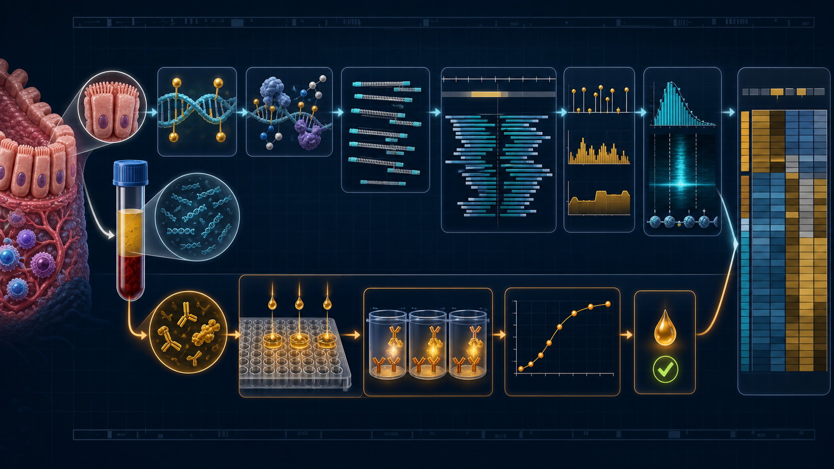
Colitis at molecular resolution
New joiner briefing
2026-09-11
Thirty minutes, five acts
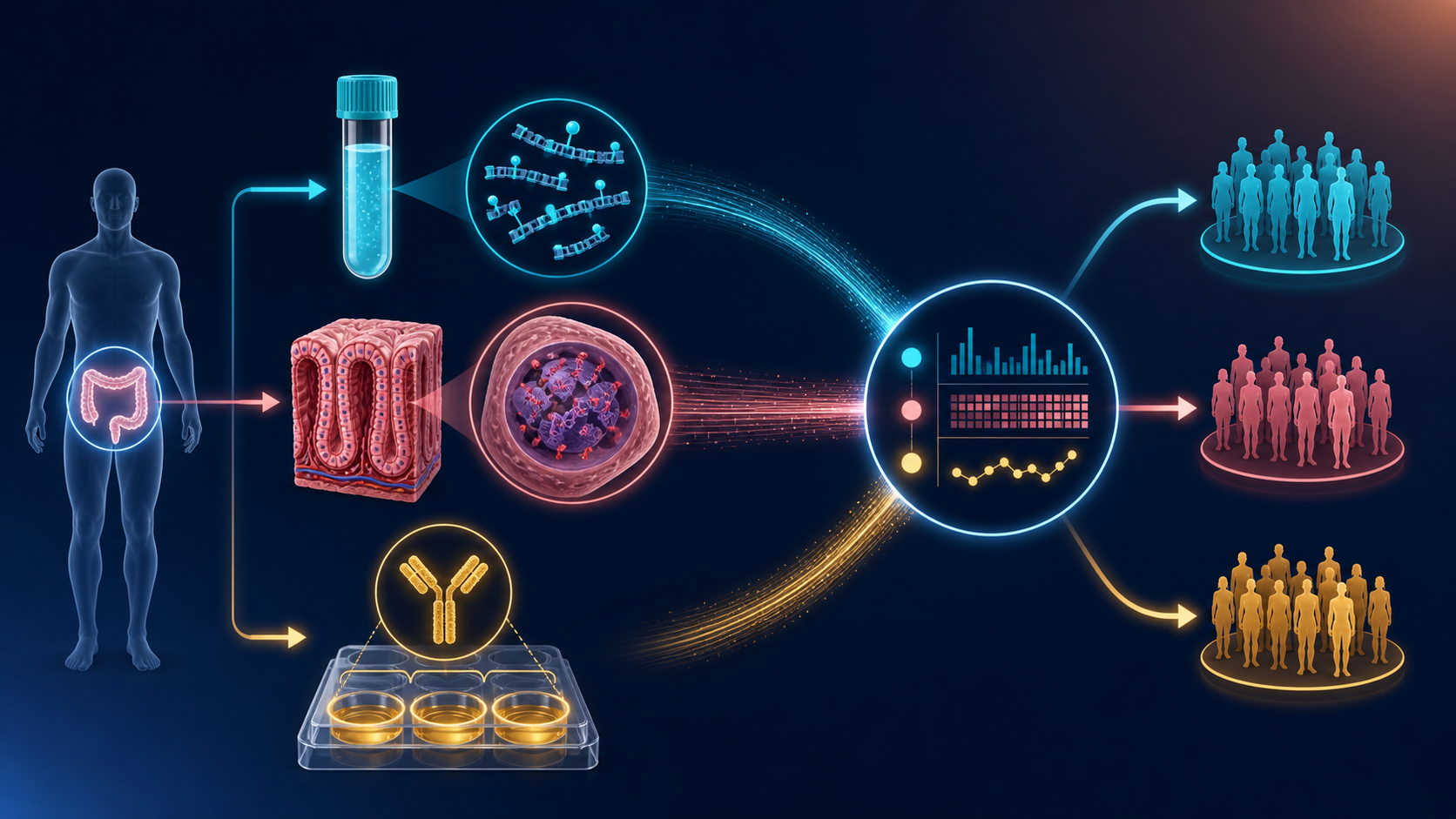
English-only briefing for new joiners. Gene names, enzymes, and software stay in their published form. Press s for timed notes. After each check, say the answer before you click.
The hour
- Clinical problem — 6 min
- Two molecular languages — 8 min
- This study — 5 min
- Lab and software — 8 min
- How you start — 3 min
Pause 20 seconds: who is wet-lab, who is computational, who is both?
Colombia, 2024: the cancer backdrop
2 minAct 1 · Clinical problem
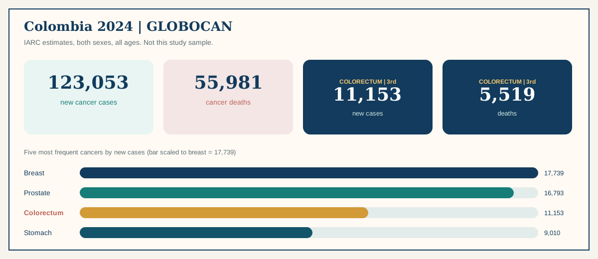
IARC estimates, both sexes, all ages. These are national figures, not this project’s sample. They explain why a Colombian colitis–CRC study exists.
Ulcerative colitis is mucosal, chronic, and not Crohn’s
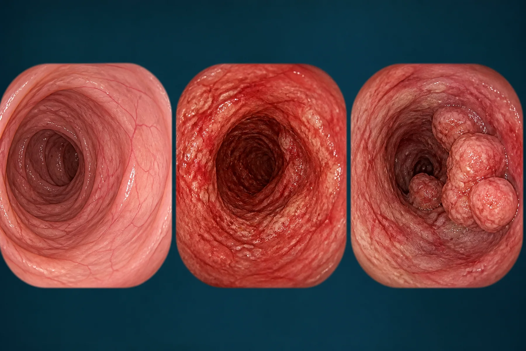
Ulcerative colitis (UC) is chronic inflammation of the colonic mucosa. Blood in stool, diarrhea, urgency, and abdominal pain are common. Extent and activity vary.
What it is
Continuous mucosal inflammation starting from the rectum in typical disease. Relapsing course.
What it is not
Not Crohn’s. This protocol excludes Crohn’s, prior colonic surgery, and contraindication to colonoscopy or biopsy.
Why eight years appears
AGA/ECCO surveillance often starts ~8–10 years after onset, or earlier with primary sclerosing cholangitis. That is a surveillance threshold, not a clock that says cancer arrives at year eight.
Progression is possible. It is not the default path.
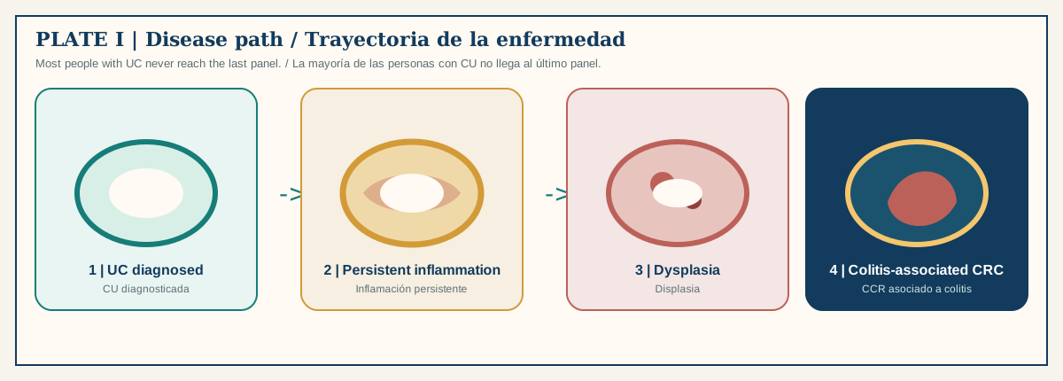
Most people with UC never enter dysplasia or colitis-associated CRC. Colonoscopic surveillance remains the clinical backbone. This project does not replace it. It asks whether blood and tissue molecules add a second, biological layer.
The research layer
Paired plasma cfDNA, colonic tissue methylation, and serum sIgA — read together, not as a diagnostic test.
Check 1
Most people with ulcerative colitis develop colorectal cancer.
False. Long-standing extensive UC raises risk. Most patients never enter dysplasia or colitis-associated CRC. Surveillance exists because cancer is possible, not because it is typical.
DNA methylation is a chemical mark, not a mutation
3 minAct 2 · Two molecular languages
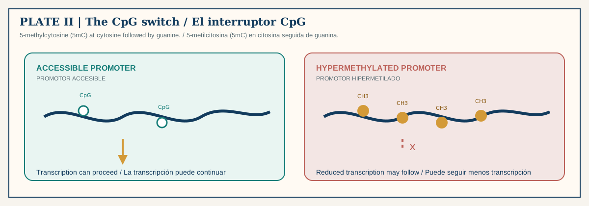
A sequencer reads C whether or not a methyl group is attached. The biology lives in that extra mark: 5-methylcytosine (5mC), usually at a CpG (cytosine then guanine on the same strand).
hypermethylation = more 5mC hypomethylation = less 5mC silenced = promoter 5mC + low RNA, measured together
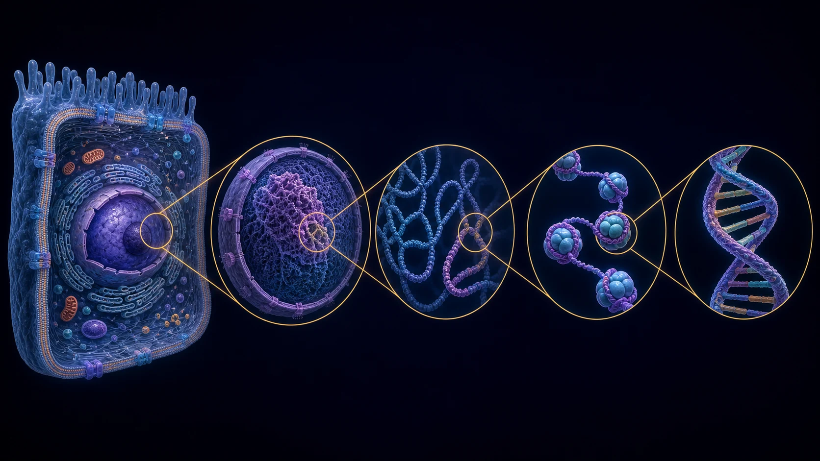
Candidate genes are hypotheses, not the disease
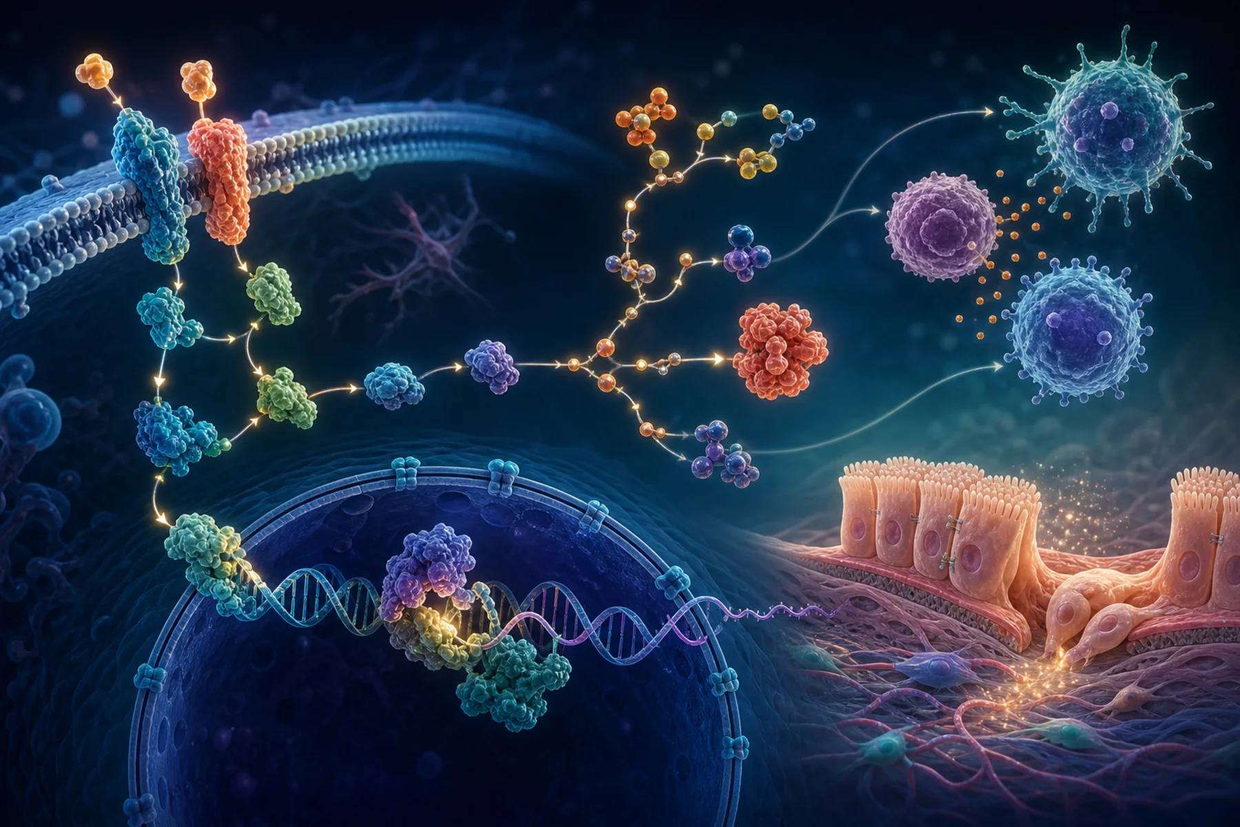
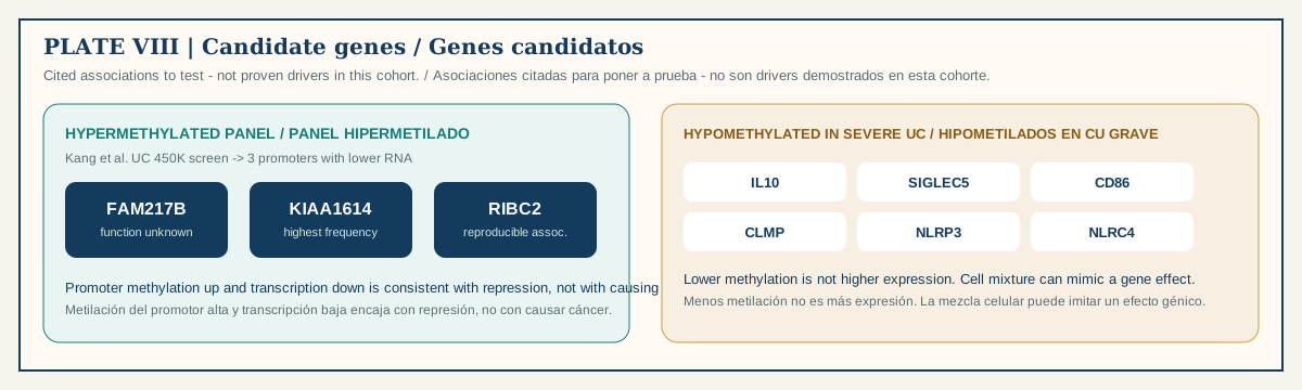
Also around UC neoplasia
CDKN2A / p16, CDH1 (E-cadherin), FOXE1 / SYNE1.
How this project must test them
Keep methylated and total counts. Use DSS. Require coherent CpGs in a region. Compare tissue with paired plasma. Adjust for inflammation and cell mixture.
Secretory IgA is a mucosal border antibody
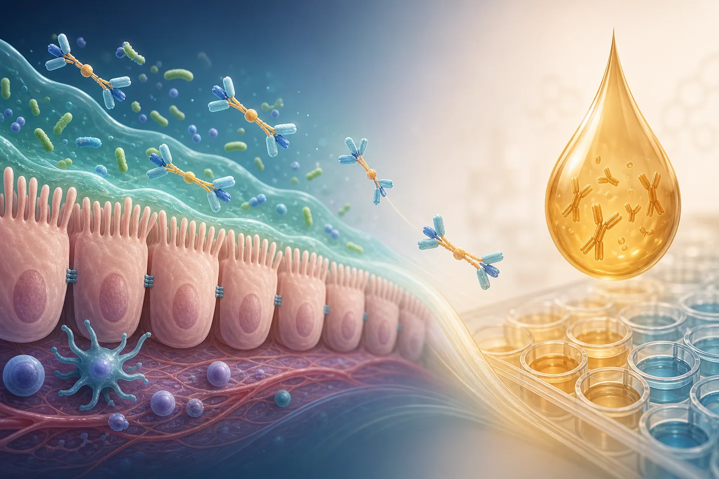
sIgA binds microbes and antigens at wet surfaces and limits epithelial contact without turning every encounter into inflammation.
This study measures serum sIgA by ELISA, calibration 1.250–0 µg/ml in the presented run (\(R^2=0.959\)). That \(R^2\) is assay development, not plate acceptance. Blanks, recovery, replicate CV, LLOQ/ULOQ, and parallelism still decide if a number is usable.
Serum is not stool, not mucus, not luminal coating. Do not report a serum value as intestinal secretion.
The two signals answer different questions

DNA methylation
Epigenetic state and cell identity. Plasma cfDNA + colonic tissue. CpG/region readout. High-dimensional. Fails under batch, cell mixture, and mixed tissue of origin.
Serum sIgA
Mucosal humoral immunity. Serum ELISA concentration. One interpretable number. Fails under infection, drugs, systemic production, and clearance.
Integration tests whether the joint pattern separates UC, CRC, and healthy groups better than either marker alone. That is still association, not a diagnostic test.
Check 2
Match the measurement to the specimen.
C. EM-seq is DNA chemistry, not an antibody assay. Tissue DNA is intentionally sheared; only native plasma fragments can support fragmentomics.
The question this grant is built to ask
2 minAct 3 · This study
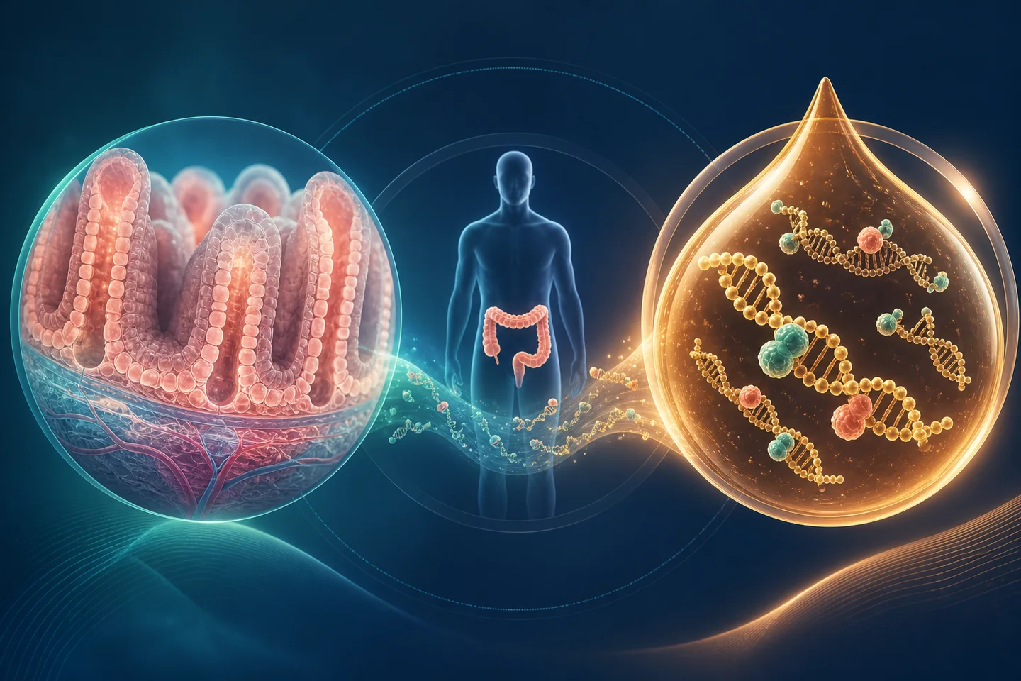
Can differential methylation in plasma cfDNA and colonic tissue, read together with serum sIgA and inflammation markers, help characterize high-risk UC relative to CRC and healthy comparators?
PCI-2024-0016 project code
18 participants planned
36 paired DNA specimens
3 clinical groups
Operational lead: Angie Alejandra Benavides Rojas. Advisor: Dr. Sandra Perdomo. Co-advisor: Dr. María Consuelo Romero. Group: InMubo.
Pairing is within a person, not between groups
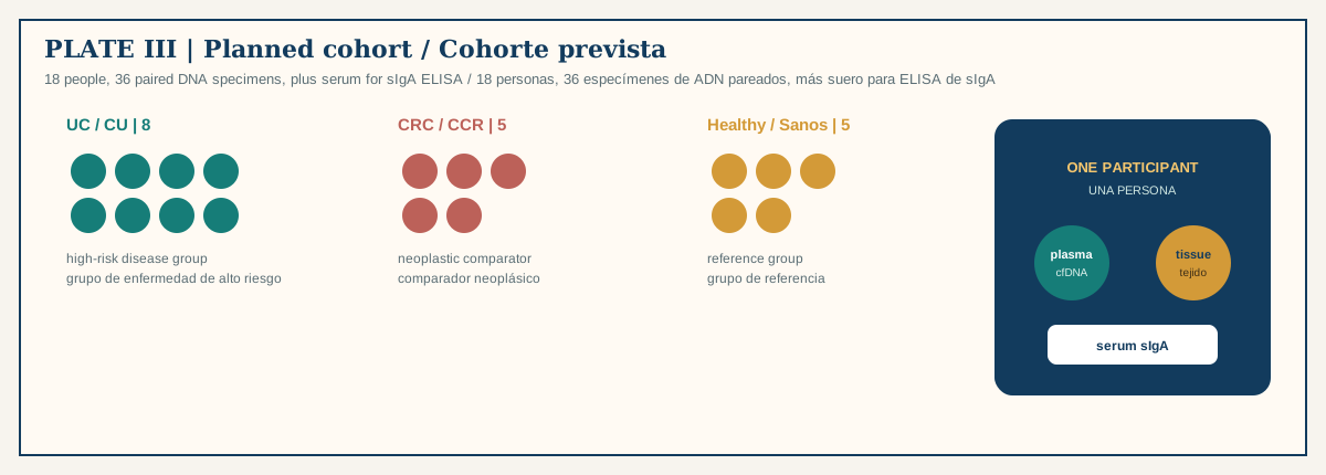
UC eligibility: confirmed UC >8 years or primary sclerosing cholangitis. Sites: Cobos Medical Center, HIC Bucaramanga, GastroAdvanced.
Ethics: Colombian Resolution 8430 of 1993; HIC Bucaramanga–Fundación Cardiovascular committee Minutes 005-2024. Public git stays de-identified. No participant-level clinical or genomic data in this repository.
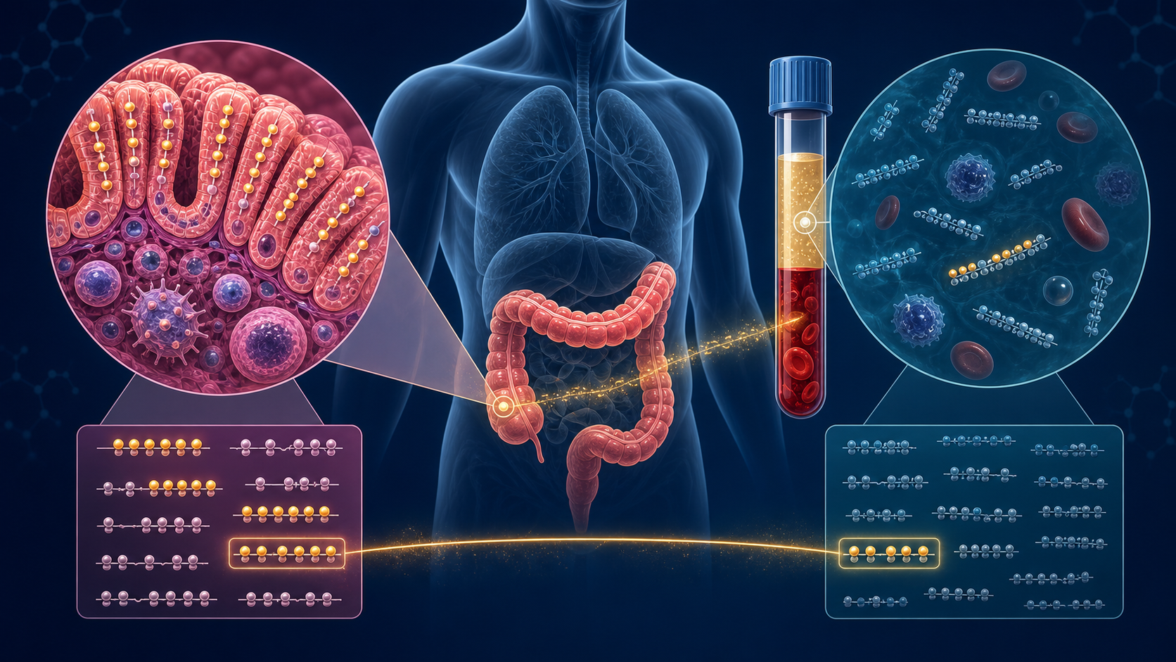
What this design can claim — and what it cannot
Signals vary together in these 18 people. This design can test that.
A model separates predefined groups in held-out data — only with locked features and honest resampling.
Predicts future progression. Needs longitudinal follow-up. Not this cross-section.
Using the model improves care versus colonoscopic surveillance. Not in scope.
Blood, biopsy, enzymes, sequencer, plate
3 minAct 4 · Tools
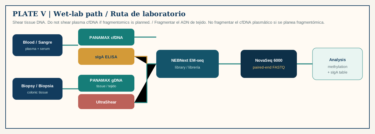
| Stage | Instrument | Stop/go |
|---|---|---|
| Plasma cfDNA | PANAMAX 48 | Yield without group-biased failure? |
| Tissue gDNA | PANAMAX 48 → UltraShear | Fragmentation documented? |
| Methylome library | NEBNext EM-seq | Conversion controls pass? |
| Sequencing | NovaSeq 6000, paired-end | Depth and identity usable? |
| sIgA | Serum ELISA | Replicates in range? |
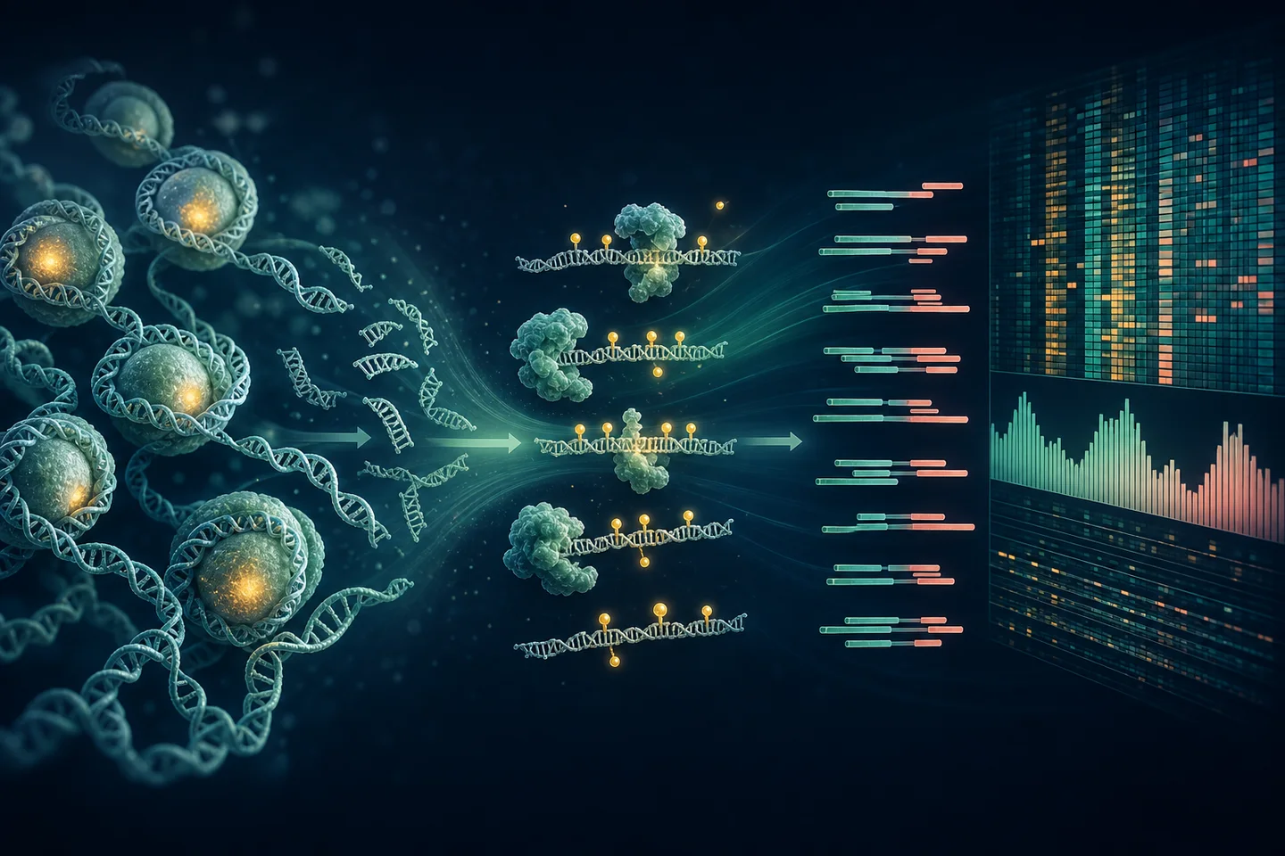
Why EM-seq instead of bisulfite
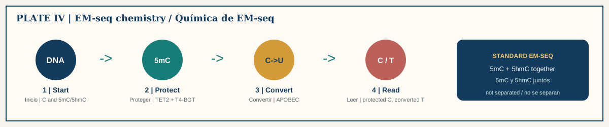
Bisulfite converts unmodified cytosines but damages DNA. Plasma cfDNA is already scarce and short. EM-seq uses TET2 + T4-BGT to protect 5mC/5hmC, then APOBEC to deaminate unmodified C, which sequences as T.
Standard EM-seq reports 5mC and 5hmC together. Write “modified cytosine” unless a second assay splits them.
Fragmentomics is a second layer on the same plasma library
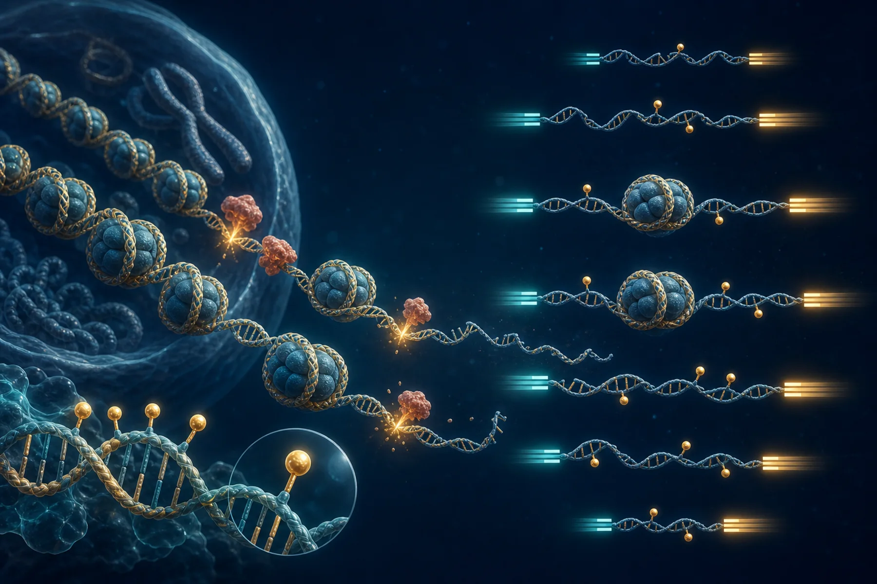
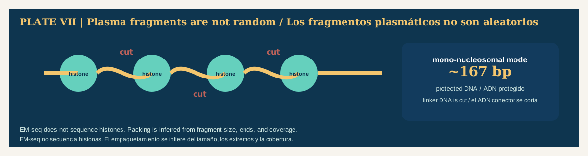
From one paired-end plasma BAM you may recover fragment length, ends, end motifs, and regional coverage. Histones are not sequenced; packing is inferred.
This is a feasibility extension. It dies if plasma was sheared, insert sizes were not retained, depth is too low, or conversion bias is uncontrolled.
From FASTQ to a region you can name

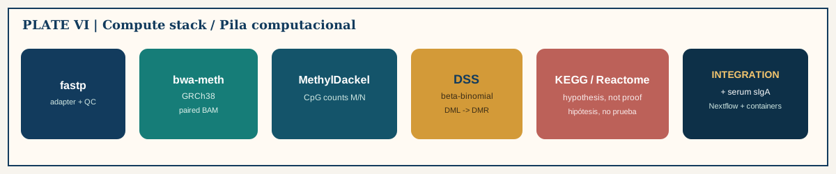
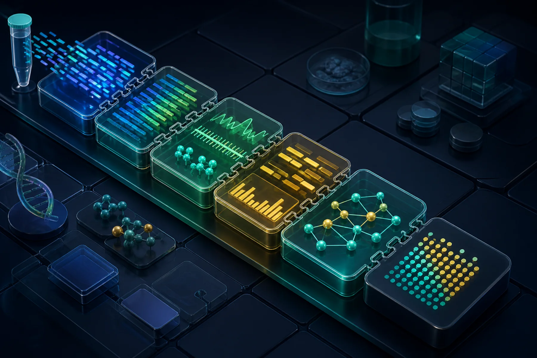
At one CpG the data are methylated reads \(M\) out of informative reads \(N\). 50% of 2 reads is not 50% of 100. DSS uses a beta-binomial model, then joins neighboring CpGs into DMRs.
Interpretation limit
KEGG/Reactome enrichment is a structured hypothesis. Record mapping rule, background universe, database version, and FDR.
Check 3
With 18 participants, the defensible near-term product is:
B. High-dimensional classifiers, prognosis, and clinical use need independent longitudinal validation. Galleri is not validated for UC progression surveillance.
Your first week in this repository
2 minAct 5 · How you start

The kernel name project-python is required even for prose-only pages.
Do not commit
Participant-level clinical or genomic data, identifiers, consent forms, linkage keys.
Freeze contract
After any edit to an R document, re-render locally and commit _freeze/. CI has no R packages.
Done today means a candidate signature, not a test
Read, in this order: study design, EM-seq, sIgA, three data streams, candidate genes, statistics, contribute.
Then take the 30-question quiz.
Closing line: this is a candidate epigenetic–immune signature with transparent uncertainty — not a diagnostic test.