flowchart LR
  P["One participant"] --> T["Colonic tissue"]
  P --> B["Blood"]
  T --> TM["Local tissue<br/>methylation"]
  B --> CF["Plasma cfDNA"]
  B --> S["Serum sIgA +<br/>inflammation"]
  CF --> PM["cfDNA methylation"]
  CF --> FR["Fragment length, ends,<br/>and coverage"]
  TM --> C["Within-person<br/>triangulation"]
  PM --> C
  FR --> C
  S --> C
Tissue versus plasma methylation sequencing: why compare both?
study design
methylation
liquid biopsy
tissue of origin
colitis
Tissue is the close-up; plasma is the blood-borne mixture. Pairing them reveals which colon methylation signals might become accessible biomarkers.
Two specimens answer different questions
Colonic tissue shows molecular change where disease occurs. Plasma cell-free DNA offers a less-invasive but mixed signal from cells throughout the body. Studying both within the same participant creates a bridge between anatomical relevance and potential clinical accessibility.
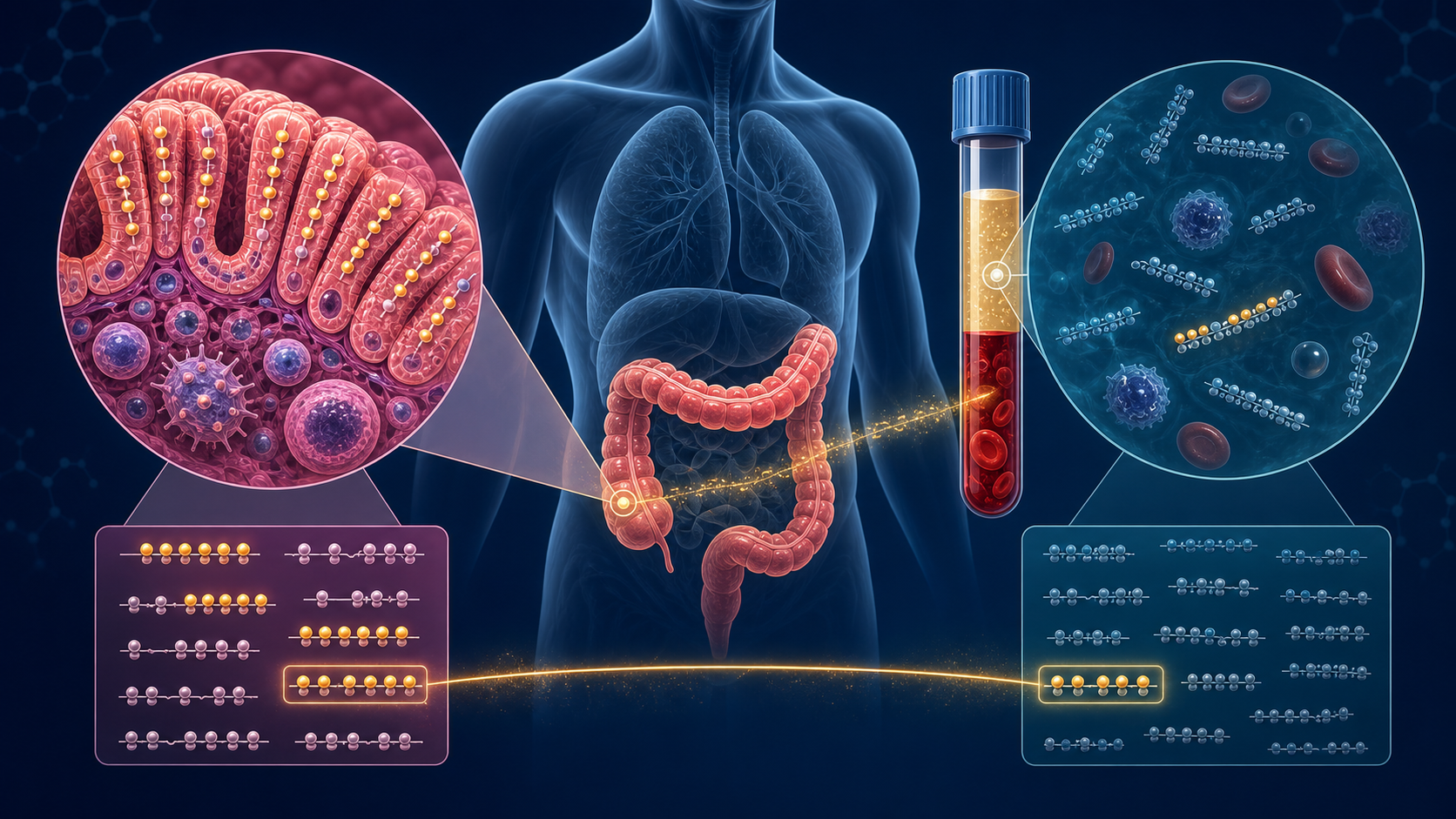
Think of tissue as a close-up photograph of one neighborhood and plasma as a bag of postcards arriving from the whole body. The close-up makes local changes easy to see. The postcards are easier to collect repeatedly, but most do not come from the colon. Comparing them asks whether a recognizable part of the local pattern survives in circulation.
The benefit in one table
| If we sequence… | We learn… | Main limitation |
|---|---|---|
| Tissue only | What is changing in the affected mucosa, at strong signal-to-noise | A biopsy is invasive, local, heterogeneous, and hard to repeat |
| Plasma only | What is detectable through a blood draw and may reflect the whole-body disease state | Colon-derived cfDNA may be rare and diluted by blood-cell DNA |
| Paired tissue + plasma | Which local methylation changes cross into blood in the same person | Small paired cohorts discover candidates; they do not validate a screening test |
The key gain is not simply “twice as much data.” Pairing gives each plasma signal a matched anatomical reference. It helps separate three explanations: a colon-linked signal that reaches blood, a local signal too diluted to observe in plasma, or a plasma signal driven by blood cells or systemic inflammation.
ImportantWhat this design can honestly claim
Paired sequencing can discover a tissue-informed plasma fingerprint. Demonstrating a classifier requires independent held-out samples; demonstrating early detection requires pre-diagnostic or prospective screening samples.
What published studies teach us
Four studies make the rationale concrete:
- A human cell-type methylation atlas showed why methylation can trace cfDNA back to contributing cell types. In a controlled mixing experiment, colon-cancer plasma remained detectable after dilution to a 3% mixture—proof of principle, not a clinical limit of detection (Moss, Magenheim, Neiman, and others, 2018).
- A plasma methylome study showed that broad, tissue- and cancer-associated methylation patterns can support tumour detection and classification even when input DNA is low (Shen, Singhania, Fehringer, and others, 2018).
- A 2024 colorectal study began with tumour-versus-adjacent-tissue DMR discovery, filtered candidates against leukocyte and cfDNA evidence, then evaluated plasma markers. Its three-DMR combination achieved AUC 0.763, 64.21% sensitivity, and 78.38% specificity in 95 CRC cases and 74 controls—promising, but also a useful reminder that tissue signal does not transfer perfectly into plasma (Long, Gao, Han, and others, 2024).
- A direct comparison in clinically relevant samples found that patient-matched cfDNA usually resembled normal samples more than tumour tissue, while some tumour-hypermethylated regions were still visible in plasma. The same study found enzymatic methylation sequencing preserved library complexity better than bisulfite conversion, supporting EM-seq for fragile, low-input cfDNA (Nuttall, Karl, Burke, and others, 2025).
Together, these papers support a disciplined sequence: use tissue to discover and interpret, use plasma to test accessibility, explicitly model leukocyte/background contributions, and reserve diagnostic claims for external validation.
How this connects to our study
The planned study includes 18 participants across three comparison groups—high-risk ulcerative colitis, colorectal cancer, and healthy comparators—with plasma cfDNA and colonic tissue collected from each participant. Serum sIgA and inflammatory measurements provide a separate immune data stream.
Pairing matters because every participant acts as a partial biological reference for themselves. Tissue–plasma comparisons are less exposed to between-person differences than comparisons made from unrelated tissue and blood cohorts, although age, sex, treatment, inflammation, cell composition, and technical batch still require attention.
What tissue contributes
Colonic biopsy DNA is anatomically anchored. A methylation difference in tissue can therefore be connected more directly to the affected mucosa than a difference detected only in plasma.
Tissue helps the project:
- identify CpGs and DMRs associated with UC or CRC tissue biology;
- compare the direction and magnitude of changes across clinical groups;
- nominate genes and pathways involving epithelial barrier function, inflammation, immune regulation, and carcinogenesis; and
- create a participant-matched reference against which plasma signals can be evaluated.
Tissue is not a perfect ground truth. A biopsy contains mixtures of epithelial, stromal, and immune cells, samples only a small anatomical area, and may be affected by inflammation, histology, treatment, and sampling depth.
What plasma contributes
Plasma cfDNA is accessible without repeatedly biopsying the colon, but it is a mixture of molecules released by blood cells and multiple tissues. Its value is translational: if a tissue-associated methylation or fragment pattern is also detectable in plasma, it may eventually support less-invasive monitoring.
Plasma adds three kinds of evidence:
- Methylation: modified-cytosine patterns that may reflect contributing cell types.
- Fragment size: short/long ratios and mono- or di-nucleosomal patterns linked to chromatin packaging and DNA release.
- Fragment position and ends: coverage around regulatory regions, genomic end coordinates, and end motifs influenced by chromatin accessibility and nuclease activity (Lo, Han, Jiang, and Chiu, 2021).
Can fragmentomics separate genes?
Fragmentomics does not physically sort genes into separate tubes. It creates genomic features that can help distinguish the activity or contribution of genomic regions and cell types.
For example, paired-end reads can be summarized around transcription start sites (TSSs). Because nucleosome positioning differs around active and inactive regulatory regions, coverage and fragment-end patterns may provide an indirect footprint of gene regulation. Fragment lengths and ends can also be aggregated over tissue-specific methylation regions or gene sets.
| Question | Fragmentomic operation | Resulting evidence |
|---|---|---|
| Is a regulatory region represented differently? | Compare normalized coverage and ends around its TSS or enhancer | Region-level footprint |
| Are fragments associated with a tissue-linked methylation signature? | Intersect fragments with pre-specified DMRs/reference regions | Candidate tissue contribution |
| Does fragment length vary with methylation? | Stratify molecule or region summaries by length and methylation level | Joint epigenetic–physical pattern |
| Do groups show different cleavage profiles? | Compare end coordinates or motif frequencies with batch-aware models | Candidate nuclease/chromatin signal |
| Are related genes changing coherently? | Aggregate tested genes into KEGG/Reactome pathways | Pathway-level hypothesis |
These analyses can prioritize genes or pathways; they do not prove that a gene is expressed. Direct transcriptional confirmation would require RNA-based measurements or another expression assay.
Four possible tissue–plasma patterns
| Observation | Possible interpretation | Next check |
|---|---|---|
| Same methylation direction in tissue and plasma | Candidate colon-linked signal detectable in circulation | Rule out blood-cell composition and batch; replicate externally |
| Strong tissue signal, weak plasma signal | Local biology may be diluted below plasma detection | Examine depth, cfDNA fraction, region coverage, and assay sensitivity |
| Plasma signal without a tissue match | Signal may originate outside the sampled biopsy or from another cell type | Test leukocyte/systemic references and biopsy heterogeneity |
| Opposite tissue and plasma directions | Mixture, sampling, technical, or biological differences may dominate | Audit QC and model cell composition before interpretation |
Agreement is informative, but disagreement is not automatically failure. It can reveal dilution, tissue heterogeneity, systemic inflammation, or a missing tissue-of-origin component.
Why add sIgA as a second biological axis?
Methylation and fragmentomics describe DNA released from cells. Serum sIgA and inflammatory markers describe an immune context. These sources are complementary rather than technical replicates.
flowchart TD
  E["Epigenetic evidence<br/>tissue + plasma methylation"] --> H["Candidate disease-state<br/>hypothesis"]
  F["Physical evidence<br/>plasma fragmentomics"] --> H
  I["Immune evidence<br/>sIgA + inflammation"] --> H
  H --> V{"Do independent biological<br/>layers agree?"}
  V -->|Yes| R["Prioritize for replication"]
  V -->|Partly| X["Refine cell-of-origin<br/>and confounding model"]
  V -->|No| N["Report uncertainty;<br/>do not force a signature"]
If a candidate methylation pattern appears in tissue and plasma and also varies coherently with immune or inflammatory measurements, the finding gains biological triangulation. Because these assays have different error structures, agreement is harder to explain by one isolated technical artifact.
However, two sources do not automatically confirm disease development. They can:
- strengthen evidence for a cross-sectional disease-state difference;
- help distinguish local colon biology from systemic signals;
- reveal whether immune state explains part of the epigenetic association; and
- prioritize a small set of candidate markers for longitudinal testing.
What would confirm progression?
Progression is a change over time. Confirming that a fingerprint tracks development from UC toward dysplasia or colitis-associated CRC would require:
- repeated tissue, plasma, and immune measurements where ethically and clinically feasible;
- dated outcomes from surveillance colonoscopy and pathology;
- a signature locked before outcome evaluation;
- models that preserve within-participant correlation and account for treatment and inflammation;
- an independent longitudinal cohort; and
- evidence that the combined signature adds information beyond established clinical surveillance.
The current study can establish feasibility, estimate cross-sectional contrasts, and nominate a tissue-informed plasma fingerprint. It cannot establish temporal progression on its own.
ImportantDesign advantage in one sentence
Paired tissue anchors the biology, plasma tests accessibility and tissue-of-origin clues, and sIgA supplies an independent immune context—together producing a stronger, more falsifiable hypothesis than any one specimen or assay alone.
Explore the full study design → · See how differential methylation is tested → · Follow the sequencing pipeline → · Understand the sIgA rationale →
References
Joshua Moss, Judith Magenheim, Daniel Neiman, and others. (2018). Comprehensive human cell-type methylation atlas reveals origins of circulating cell-free DNA in health and disease. Nature Communications. https://doi.org/10.1038/s41467-018-07466-6.
Shu Yi Shen, Rajat Singhania, Gordon Fehringer, and others. (2018). Sensitive tumour detection and classification using plasma cell-free DNA methylomes. Nature. https://doi.org/10.1038/s41586-018-0703-0.
Zhiping Long, Yu Gao, Zhen Han, and others. (2024). Discovery and validation of methylation signatures in circulating cell-free DNA for the detection of colorectal cancer. Biomolecules. https://doi.org/10.3390/biom14080996.
Barrett Nuttall, Daniel L. Karl, Kathleen Burke, and others. (2025). Comprehensive comparison of enzymatic and bisulfite DNA methylation analysis in clinically relevant samples. Clinical Epigenetics. https://doi.org/10.1186/s13148-025-01959-0.
Y. M. Dennis Lo, Diana S. C. Han, Peiyong Jiang, and Rossa W. K. Chiu. (2021). Epigenetics, fragmentomics, and topology of cell-free DNA in liquid biopsies. Science. https://doi.org/10.1126/science.aaw3616.