flowchart LR
  C["Cell nucleus"] --> N["DNA wrapped around<br/>histone nucleosomes"]
  N --> D["Cell death +<br/>nuclease cleavage"]
  D --> F["Protected cfDNA<br/>fragments in plasma"]
  F --> L["Length + ends +<br/>coverage footprints"]
  F --> M["Methylation state<br/>along the fragment"]
  L --> I["Joint biological<br/>interpretation"]
  M --> I
Sequencing, methylation & fragmentomics
What EM-seq measures, how the genomics pipeline works, and why cfDNA fragments reflect chromatin
What is actually sequenced?
The sequencer reads both ends of short DNA molecules in an EM-seq library. For each paired-end plasma molecule, the analysis may recover:
| Observable | Derived from | Biological layer |
|---|---|---|
| Read sequence and genomic position | Bases and alignment to GRCh38 | Genomic origin of the molecule |
| Cytosine state | Enzymatic conversion followed by sequencing | 5mC/5hmC protection versus unmodified cytosine conversion |
| Fragment length | Distance and orientation of a properly paired alignment | Size distribution and nucleosomal periodicity |
| Fragment ends | Left/right genomic coordinates and terminal sequence motifs | Chromatin- and nuclease-associated cleavage patterns |
| Regional coverage | Counts of overlapping fragments | Genome-wide representation and footprints near regulatory regions |
Fragmentomics does not sequence a separate molecule type. It reuses positional and physical information carried by paired-end reads from native plasma cfDNA. Tissue genomic DNA is deliberately sheared for library preparation, so its insert lengths describe the laboratory fragmentation step and should not be interpreted as native tissue fragmentomics.
Why histones matter
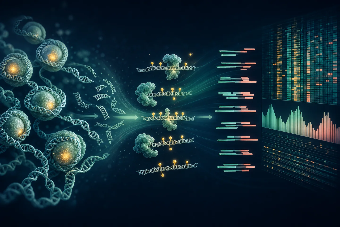
DNA in the nucleus is wrapped around histone proteins to form nucleosomes. During cell death and DNA release, nucleosome-protected DNA is less accessible to nucleases than linker DNA. Plasma cfDNA therefore often shows a mono-nucleosomal mode near 167 bp and longer multiples, plus coverage and end-position patterns related to chromatin organization (Lo, Han, Jiang, and Chiu, 2021).
Histones are not directly sequenced by EM-seq. Their organization is inferred indirectly from patterns in DNA fragment size, positions, and coverage. This inference can be altered by collection delay, nuclease activity, extraction, end repair, conversion, PCR, sequencing depth, and mapping.
How methylation and fragmentation connect
Methylation is associated with chromatin state, and cleavage near CpGs can differ with methylation configuration. FRAGMA demonstrated methylation-aware CpG cleavage profiles (Zhou, Kang, Jiang, and others, 2022), while FinaleMe used fragment length, coverage, and CpG position within a fragment to infer methylation from plasma WGS (Liu, Reed, Lo, Choudhury, and others, 2024). These papers justify testing linked features, but their trained models are not automatically valid for this EM-seq protocol or UC cohort.
NEBNext EM-seq wet-lab logic
NEBNext Enzymatic Methyl-seq avoids the harsh bisulfite reaction and supports low-input, fragmented DNA (Vaisvila, Ponnaluri, Sun, and others, 2021). In simplified terms:
- Prepare the DNA library. Preserve native plasma cfDNA; shear tissue DNA in a controlled, documented step.
- Protect modified cytosines. TET2 oxidation and T4-BGT chemistry protect 5mC and 5hmC from the later deamination step.
- Convert unmodified cytosines. APOBEC deaminates unmodified cytosine so it is read as thymine after amplification and sequencing.
- Sequence paired ends. Illumina reads retain methylation information and, for plasma, the paired alignment needed to estimate insert length and ends.
NoteWhat the assay reports
Standard EM-seq readout generally represents protected 5mC and 5hmC together rather than separating them. Interpret it as modified-cytosine signal unless an additional assay distinguishes the two.
Genomics pipeline
flowchart TD
  R["Paired FASTQ"] --> Q["fastp<br/>adapter + quality QC"]
  Q --> A["bwa-meth<br/>alignment to GRCh38"]
  A --> B["Coordinate-sorted BAM<br/>pair + insert QC"]
  B --> MC["MethylDackel<br/>CpG methylation calls"]
  MC --> DS["DSS<br/>DML/DMR models"]
  DS --> PA["Gene mapping +<br/>KEGG/Reactome pathways"]
  B --> FR["Fragment feature table<br/>lengths, ends, TSS coverage"]
  MC --> JM["Methylation × length<br/>joint summaries"]
  FR --> JM
  PA --> IN["Integrated model"]
  JM --> IN
  S["sIgA + inflammation<br/>+ clinical covariates"] --> IN
Protocol-by-protocol guides
| Stage | Guide |
|---|---|
| 18 consented participants · three groups | Cohort, consent, and study manifest |
| Blood → plasma + serum | Blood processing and aliquots |
| Colonic biopsy → prepared tissue | Colonic biopsy preparation |
| PANAMAX 48 · plasma | Automated cfDNA extraction |
| PANAMAX 48 · tissue | Automated genomic-DNA extraction |
| UltraShear | Tissue-DNA fragmentation |
| NEBNext EM-seq | Methylation library preparation |
| NovaSeq 6000 | Paired-end sequencing |
| Serum sIgA | ELISA processing and data |
Minimum outputs and checks
| Stage | Required output | Stop/go question |
|---|---|---|
| FASTQ QC | Read counts, base quality, adapter content, duplication indicators | Is the library usable without differential quality by group? |
| Alignment | Mapping rate, proper-pair rate, duplicates, conversion controls, insert sizes | Are positions, methylation calls, and native plasma sizes credible? |
| Methylation | Coverage distribution, conversion performance, CpG calls | Is coverage sufficient and comparable for DML/DMR modeling? |
| Fragmentomics | Length distribution, short ratio, nucleosomal peaks, ends, TSS coverage | Was native fragment structure preserved and is depth adequate? |
| Integration | Locked feature table with batch and clinical covariates | Can uncertainty be estimated without feature leakage or overfitting? |
Pathway interpretation
After FDR-controlled DML/DMR analysis, mapped genes can be summarized with KEGG and Reactome. The report should retain the CpG/DMR-to-gene rule, tested-region background, database version, effect direction, overlap size, and adjusted p-value. Enrichment is a structured hypothesis about biological context—not direct evidence that a pathway is active.
Learn what KEGG contributes → · Explore Reactome’s reaction hierarchy →
ImportantCritical boundary
With 18 planned participants, the defensible target is a quality-controlled feasibility analysis and a small set of candidate features. High-dimensional classifiers, prognosis claims, and clinical use require independent longitudinal validation.
See the complete experimental roadmap → · Read the literature review →
Prefer a visual introduction? Read “What is enzymatic methyl sequencing?” →
Then explore “What is fragmentomics?” →
See how the analysis software evolved → · Understand beta-binomial and regional statistics →
References
Y. M. Dennis Lo, Diana S. C. Han, Peiyong Jiang, and Rossa W. K. Chiu. (2021). Epigenetics, fragmentomics, and topology of cell-free DNA in liquid biopsies. Science. https://doi.org/10.1126/science.aaw3616.
Qing Zhou, Guannan Kang, Peiyong Jiang, and others. (2022). Epigenetic analysis of cell-free DNA by fragmentomic profiling. Proceedings of the National Academy of Sciences. https://doi.org/10.1073/pnas.2209852119.
Yaping Liu, Sarah C. Reed, Christopher Lo, Atish D. Choudhury, and others. (2024). FinaleMe: Predicting DNA methylation by the fragmentation patterns of plasma cell-free DNA. Nature Communications. https://doi.org/10.1038/s41467-024-47196-6.
Romualdas Vaisvila, Vamsi K. C. Ponnaluri, Zhe Sun, and others. (2021). Enzymatic methyl sequencing detects DNA methylation at single-base resolution from picograms of DNA. Genome Research. https://doi.org/10.1101/gr.266551.120.