flowchart TD
  subgraph DNA[EM-seq branch]
    T[Tissue DNA<br/>controlled shearing] --> L[EM-seq library]
    P[Native plasma cfDNA] --> L
    L --> Q[Paired FASTQ<br/>fastp + FastQC/MultiQC]
    Q --> A[bwa-meth<br/>GRCh38 alignment]
    A --> B[Sorted/indexed BAM<br/>read groups + duplicate/QC]
    B --> MB[M-bias + conversion<br/>diagnostics]
    MB --> C[MethylDackel<br/>methylated/total CpG counts]
    C --> BS[BSseq/count matrices]
    BS --> DSS[DSS DML/DMR contrasts]
    B --> F[Plasma-only native<br/>fragment features]
  end

  subgraph ELISA[sIgA branch]
    S[Serum] --> W[Standards, blanks,<br/>controls + replicate wells]
    W --> OD[Blank-corrected OD]
    OD --> CAL[Calibration curve +<br/>dilution correction]
    CAL --> IG[One reviewed sIgA<br/>estimate + QC]
  end

  DSS --> J[Participant-level feature matrix]
  F --> J
  IG --> J
  X[Clinical group, inflammation,<br/>covariates + batch] --> J
From EM-seq reads to an integrated study matrix
bioinformatics
alignment
EM-seq
cfDNA
sIgA
The alignment, methylation, fragmentomics, DMR, and sIgA processing flow for a genomics audience
Scope
This is the technical handoff from paired-end EM-seq FASTQs and ELISA plate readings to analysis-ready participant features. The genomics branch uses conversion-aware alignment and retains methylated/total counts; the sIgA branch is a separate quantitative assay that joins only after its own calibration and QC.
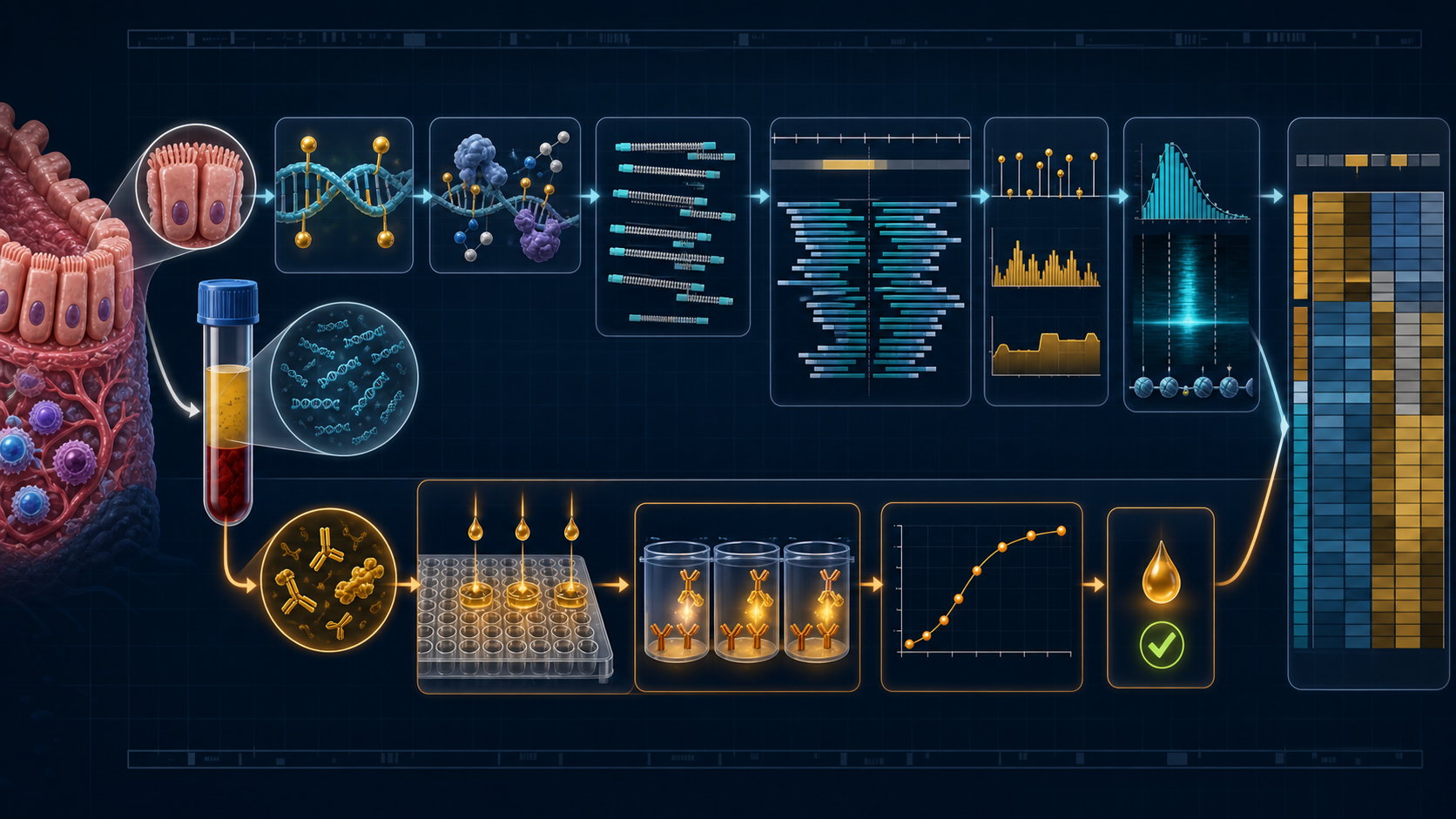
Assay topology
For each of the 18 planned participants, the design links colonic tissue DNA, plasma cfDNA, and serum sIgA. The sequencing libraries are not interchangeable:
- Tissue DNA is extracted from a heterogeneous biopsy and intentionally fragmented during library preparation. Its methylation is biologically interpretable; its insert-size distribution is largely a laboratory product.
- Plasma cfDNA arrives pre-fragmented by in-vivo release, nucleosome protection, and nuclease cleavage. If preparation preserves molecule geometry, paired alignments support methylation plus native fragmentomics.
- Serum sIgA is measured by ELISA. No sIgA sequence reads enter the genomics workflow.
Three laboratory technical replicates from a participant remain nested under that participant. They provide reproducibility and QC information, not additional biological degrees of freedom.
End-to-end flow
1. Sample sheet and identity are first-class data
Before sequence processing, a machine-readable manifest should bind participant_id, specimen, technical replicate, library, lane, read files, collection time, extraction batch, library batch, and clinical group. The participant identifier—not the library name—is the join key used in analysis.
Identity and contamination checks should be resolved before group modeling. Replicates must agree genetically where informative, and tissue/plasma labels should not be inferred from filenames alone. No clinical identifier belongs in public analysis artifacts.
2. EM-seq chemistry changes what alignment means
NEBNext EM-seq protects modified cytosines through TET2 oxidation and T4-BGT glucosylation, then APOBEC converts unmodified cytosines so they are read as thymine after amplification (Vaisvila, Ponnaluri, Sun, and others, 2021). Standard EM-seq therefore reports protected 5mC + 5hmC rather than separating them.
The aligner must tolerate conversion-induced C→T and complementary G→A differences. bwa-meth presents a BWA-MEM-based conversion-aware mapping path against GRCh38 and emits conventional SAM/BAM coordinates for downstream tools.
3. FASTQ and alignment QC
Adapter/quality trimming with fastp should produce both cleaned reads and per-library JSON/HTML metrics. The point is not maximal trimming; it is removal of demonstrated adapter/read-through and low-quality tails without erasing short cfDNA molecules.
After alignment, retain at least:
| Metric | Why it matters |
|---|---|
| total and retained read pairs | library yield and differential attrition |
| mapped and properly paired fraction | trustworthy coordinates and inserts |
| MAPQ distribution | ambiguous mapping and repetitive regions |
| duplicate fraction | independent library complexity |
| insert-size distribution by specimen | plasma nucleosomal pattern; tissue shearing QC |
| mitochondrial/off-target fraction | sample and preparation behavior |
| spike-in conversion controls | incomplete conversion or overconversion |
| read-cycle methylation/M-bias | end repair or position-specific bias |
Coordinate sorting, indexing, read-group completeness, flag statistics, and duplicate policy must be consistent across libraries. Group must not be confounded with lane or preparation batch.
4. M-bias-aware methylation extraction
MethylDackel can inspect per-cycle methylation bias and extract strand-aware cytosine calls from the BAM. End trimming parameters should be chosen from the observed M-bias profile and fixed by library class, rather than tuned to maximize group separation.
The irreducible per-CpG record is:
chr pos strand/context methylated_count unmethylated_count total_coverageCHG/CHH calls and spike-ins are useful conversion diagnostics even when biological inference is restricted to CpG sites. Filters should address MAPQ, base quality, overlapping mate double-counting, duplicates, extreme depth, blacklisted/problematic loci, strand combination, and minimum sample coverage. Every transformation should be represented in a QC report and a versioned callable-loci mask.
5. Replicates become count-aware matrices
After technical-replicate QC, valid sequencing replicates can be combined at the evidence level where the laboratory design supports pooling:
\[M_{gi}=\sum_r M_{gir},\qquad N_{gi}=\sum_r N_{gir}.\]
This preserves the difference between, for example, 5/10 and 50/100. Averaging replicate beta values would discard that precision structure. If technical batches are scientifically important or replicates are not exchangeable, keep them explicit in a hierarchical model instead.
The analysis object should retain genomic ranges, \(M\), \(N\), sample metadata, callable status, technical provenance, and specimen type. Tissue and plasma can share coordinates without being forced into the same coverage filter.
6. Differential methylation and regions
For raw EM-seq counts, DSS supplies a beta-binomial, dispersion-shrunk route from CpGs to DMLs and DMRs. Pre-specified contrasts are UC versus healthy, CRC versus healthy, and CRC versus UC, with a parsimonious design for batch and defensible covariates.
DMR output should retain:
- genomic interval and number of contributing CpGs;
- direction and magnitude of methylation difference;
- test statistic, raw p-value, and FDR;
- coverage/callability by group and specimen;
- gene, promoter, enhancer, CpG-island, and pathway annotations; and
- whether direction agrees in matched tissue and plasma.
The limma-versus-DSS comparison explains why coverage belongs in the primary likelihood and includes a runnable real-data example.
7. Plasma fragmentomics branches from BAM—not from tissue inserts
Properly paired, primary plasma alignments can yield template length, left/right end coordinates, end motifs, window coverage, and TSS-centered footprints. Deduplication, clipping, MAPQ, read overlap, and conversion-aware sequence interpretation must be specified. End motifs observed after conversion chemistry are not automatically equivalent to motifs from ordinary WGS.
Fragment features should be aggregated before participant-level modeling: length-bin proportions, short/long ratios, nucleosomal modes, regional end counts, and normalized coverage around locked regions. Tissue insert sizes should remain library QC unless native tissue fragmentation was preserved by a distinct protocol.
8. sIgA processing is a separate quantitative pipeline
The serum assay begins with plate layout: standards, blanks, controls, participant wells, dilution, replicate, plate, and position. The presentation describes a seven-point 1.250-to-0 µg/ml development curve with reported \(R^2=0.959\); that number alone does not validate a plate.
A reproducible processing path should:
- subtract or model blank signal;
- fit the pre-specified calibration function, commonly a validated 4PL/5PL form where appropriate;
- back-calculate concentration and apply the dilution factor;
- check standard recovery, monotonicity, residuals, controls, and plate drift;
- compute replicate CV and apply pre-specified repeat/failure rules;
- flag values below LLOQ, above ULOQ, or saturated rather than silently imputing them; and
- emit one reviewed participant estimate plus replicate- and plate-level provenance.
participant_id  siga_concentration  replicate_cv  plate  censor_flag  qc
P001            0.73                0.021         A01    NONE         PASS
sIgA is then joined to methylation/fragment features by participant ID. It should not be normalized like a sequencing count matrix or treated as a surrogate for local mucosal IgA.
9. The contract between pipelines
The final integration layer should be narrow and explicit:
| Output object | Unit | Typical columns |
|---|---|---|
| methylation regions | participant × DMR | estimate, coverage, uncertainty, specimen |
| fragment summary | participant × feature | normalized count/proportion, plasma QC |
| sIgA summary | participant | concentration, CV, censor/QC flags |
| phenotype table | participant | group, inflammation, covariates, batch |
Joining all rows into one table does not remove their different error models. High-dimensional discovery occurs with count-aware methods; sIgA and fragment summaries enter association or small integrated models only after QC and dimension reduction.
ImportantExpert takeaway
The pipeline preserves the observation model at every handoff: converted paired reads become coordinate-aware methylated/total counts; only native plasma alignments become fragmentomics; ELISA wells become one calibrated sIgA estimate; and all technical replicates collapse to—or are modeled under—the participant, the biological unit.
See the complete study design → · Inspect example processed tables → · Read the ML framing → · Read the statistical framing → · Open the sequencing methods page →
References
Romualdas Vaisvila, Vamsi K. C. Ponnaluri, Zhe Sun, and others. (2021). Enzymatic methyl sequencing detects DNA methylation at single-base resolution from picograms of DNA. Genome Research. https://doi.org/10.1101/gr.266551.120.